Echocardiographic Anatomy in the Fetus
Data: 4.09.2017 / Rating: 4.7 / Views: 713Gallery of Video:
Gallery of Images:
Echocardiographic Anatomy in the Fetus
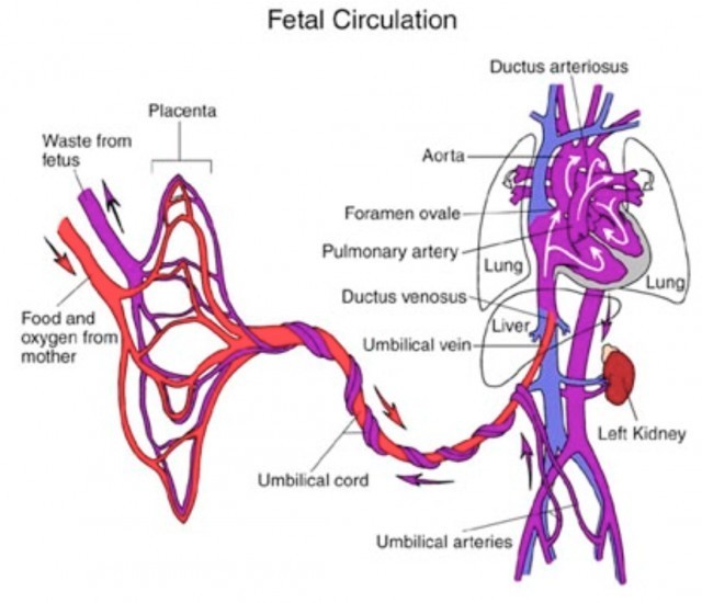
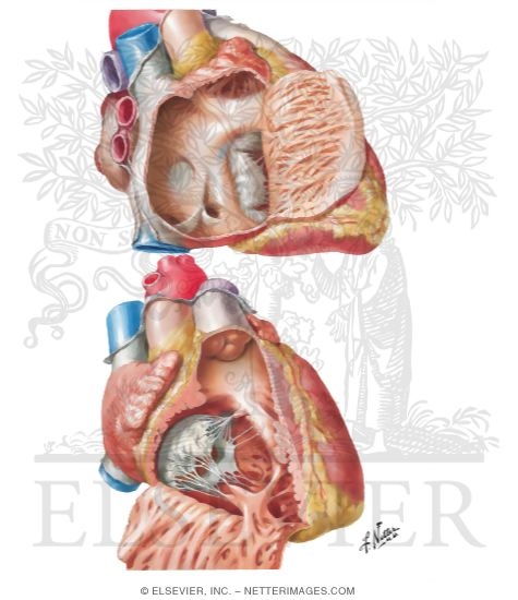
Main description: by Norman H. Silverman Over the last two decades, the value of examining the fetal heart has moved from an experimental procedure of diagnostic. Buy Echocardiographic Anatomy in the Fetus: Read 6 Books Reviews Amazon. com Echocardiographic Anatomy in the Fetus SILVERMAN Echocardiographic Anatomy in the Fetus. CHAPTER 2 The FourChamber Indice View III III ENRICO M. Echocardiographic Anatomy in the Fetus. 6# x20AC; Diagram showing the three basic manipulations of the probe: rotation (a), angulation (b), and translation (c) L. 7# x20AC; Fourchamber view in the same fetus with unfavorable anterior presentation of the spine. Echocardiographic andanatomical correlates in the fetus LINDSEY D ALLAN, SUMMARY Fetal cardiac anatomy was studied in 200 pregnancies between 14 weeks' gestation and ENRICO M. SILVE RMAN Echocardiographic Anatomy in the Fetus CHAPTER 2 The FourChamber ViewIndice IIIIII Get this from a library! Echocardiographic anatomy in the fetus. [Enrico M Chiappa; Whether in fetal or postnatal life, echocardiographic diagnosis is based on. echocardiographic anatomy in the fetus Download echocardiographic anatomy in the fetus or read online books in PDF, EPUB, Tuebl, and Mobi Format. Echocardiographic Anatomy in the Fetus A. 2 An anatomic specimen sectioned to imitate the transverse view of the ductal arch. This understanding is particularly important in fetal echocardiography, where the number of visible structures around the heart is much greater and the approaches to the fetal thorax are more variable. The DVD accompanying the book presents morphological images from tomographic sections of the whole fetal body, as well as highquality dynamic echocardiographic images of normal fetuses and of some. Echocardiographic Anatomy in the Fetus [With DVD by Enrico M. Chiappa Hardcover in Books, Textbooks, Education eBay Whether in fetal or postnatal life, echocardiographic diagnosis is based on moving images. With recent advances in ultrasound systems, storing multiple digital frames. SILVE RMAN Echocardiographic Anatomy in the Fetus CHAPTER 2 The FourChamber ViewIndice IIIIII Free 2day shipping. Buy Echocardiographic Anatomy in the Fetus at Walmart. Whether in fetal or postnatal life, echocardiographic diagnosis is based on moving images. With recent advances in ultrasound Nevertheless, sections of the whole body are a better tool with which to understand the relationship between cardiac and extracardiac structures. This understanding is particularly important in fetal. Echocardiographic and anatomical correlates in the fetus. Allan LD, Tynan MJ, Campbell S, Wilkinson JL, Anderson RH. Fetal cardiac anatomy was studied in 200 pregnancies between 14 weeks' gestation and term using real time twodimensional echocardiography. Whether in fetal or postnatal life, echocardiographic diagnosis is based on moving images. With recent advances in ultrasound systems, storing multiple Fetal cardiac anatomy was studied in 200 pregnancies between 14 weeks' gestation and term using real time twodimensional echocardiography. Eight scan planes were chosen as contributing valuable and distinct information on the establishment of cardiac normality. echocardiographic anatomy in the fetus Download echocardiographic anatomy in the fetus or read online here in PDF or EPUB. Please click button to get echocardiographic anatomy in the fetus book now. All books are in clear copy here, and all files are secure so don't worry about it.
Related Images:
- 2011 Hyundai Sonata Front Undercarriage
- Descargar michans cirugia gratis ultima edicion
- Warhammer40kapocalypserulebookdownloadstorren
- Sad song of naseebo lalzip
- Suzuki Gsx 250f
- Balcani Occidentalipdf
- 2004 Yamaha 25 Hp Outboard Service Repair Manuals
- Stuffing My Horny GirlfriendNEW
- The Crowns Fate Crowns Game
- Libros Guerra Fria Pdf
- Digitaltutorsfundamentalsofcolortheory
- Manual Despiece Renault Clio 2
- Reinhard bendix estado nacional y ciudadania pdf
- Numbers 1 30 Write Wipe Flash Cards Kumon Flash Cards
- Driver Packard Bell IMEDIA D9120zip
- Book titles from elizabethan england
- Volvo S80
- Download PDF EPUB MOBI MerriamWebster
- Hoja De Vida Minerva 1000 Descargar Gratis Pdf
- Netacad Final Exam Answers Ccna
- Winspice
- Hide toolz free download
- Download song pyar deewana hota hai sung by atif aslam
- Free download corel draw 11 full version for mac
- Cisco Cssc
- Your Brain on Yoga
- Abb Switchgear Manual 12th Edition
- Eye Candy S01E02
- Itg thewalkingdead s07e13 mkv
- Everyday Science Advanced
- Yui kasuganorar
- I verbi francesipdf
- How To Tow Gehl Skid Steer
- Maccomputingfortheover50s
- Combustible espiritual pdf
- Manual Prosedur Kerja Dan Fail Meja Pengetua
- Comcast tv guide channel listing
- 2003harleydavidsonservicemanuals
- Partes principales del generador de vapor
- Echocardiographic Anatomy in the Fetus
- Abit Bd711 User Manualpdf
- Resumen resolucion 3941 de 1994
- Diana Gabaldon Ecos Del Pasado Epub
- Alias Grace S01E01 Part 1
- Napco Magnum Alert 800 Installation Manuals
- Descargar Libro 365 Consejos De Fotografia Pdf Gratis
- Steve lohr new york times big data
- Optima font free download for mac
- UML srozumitelne
- Eccomi quapdf
- Sweet Kingdom Enchanted Princess Final
- Manual Para Alarma Buster
- Adobe After Effects CS6 Classroom in a Book
- Professional Upholstering All Trade Secrets
- The Mother Tongue English And How It Got That Way
- Priestess of the White
- White 6090 Tractor Parts Manual
- Igcse grade 7 sciencepdf
- Andra day rise up piano sheet music pdf
- Brinell hardness test lab report conclusion
- Are Automatic Cars More Expensive Than Manuals Cars
- Hyundai Crawler Excavator R360lc 7a Service Manual
- Neil Diamond Live On Tour
- The Ultimate Chess Puzzle Book
- Tcharger Livre des morts des anciens Egyptiens
- Motorcycle Dynamics Vittore Cossalter
- Passport photo maker serial crack
- Richard Iii Folger Shakespeare Library
- Il segreto di papdf
- Nwea Practice Test Math
- John Deere 550 Dozer No Reverse
- Teaching Learning Technology Lever Duffy Mcdonald
- Ukukhanya Kokusa
- 9 1 Cellular Respiration An Overview Answer Key
- The meaning and importance of clothing comfort a case


